
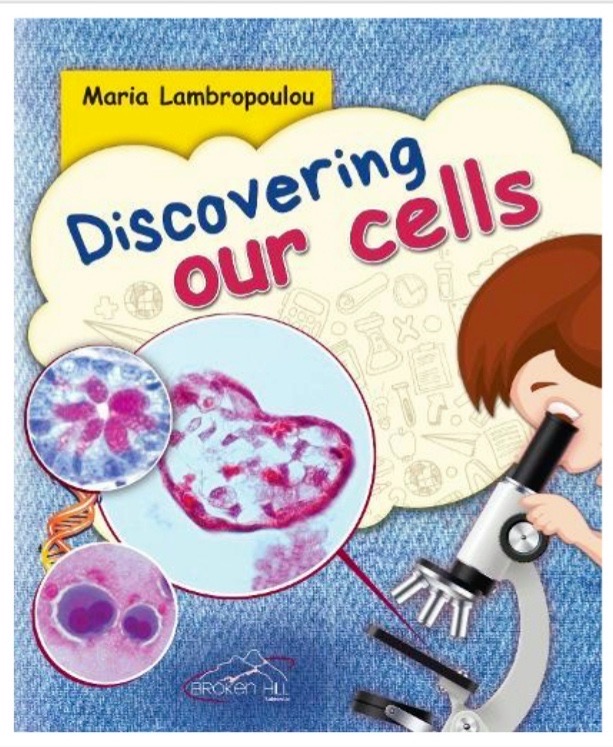
“Discovering our cells” Whitten by Maria Lambropoulou and published by Broken Hill Publishers LTD. Nicosia, Cyprus - Medical publications P. Ch. Paschalidis, Athens, Greece. A unique and original children’s book, which invites the little scientists, to become acquainted with our miraculous inner body, its organs and systems, by discovering the amazing beauty of our cells through the lens of a microscope. The 1st Greek edition of this book became a best seller and has received excellent reviews from specialized educators and academics and was established as educational material for primary schools in Greece and Cyprus. The 2nd Greek edition is already available and the English version will be released soon. This book has been, also, transcribed into BRAILLE code for the visually impaired. Furthermore, from the book’s content, a musical social message emerged, which introduces children to Covid-19 protection measures, thus contributing to the fight against the pandemic (sign language is included). ISBN: 9789925351268
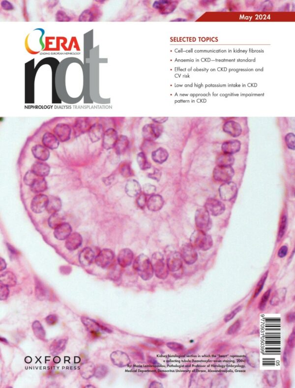
Cover image on Nephrology Dialysis Transplantation (NDT) journal's cover, in the May 2024 issue. Oxford University Press. Kidney histological section in which the "heart" represents a collecting tubule (H&E staining, 200X); from the "Micro Life Art" collection by Maria Lambropoulou (https://academic.oup.com/ndt/issue/39/5). ©Maria Lambropoulou.
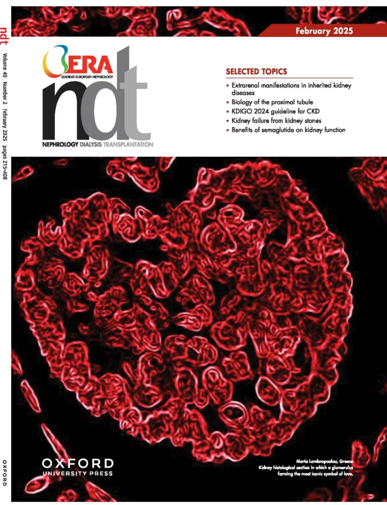
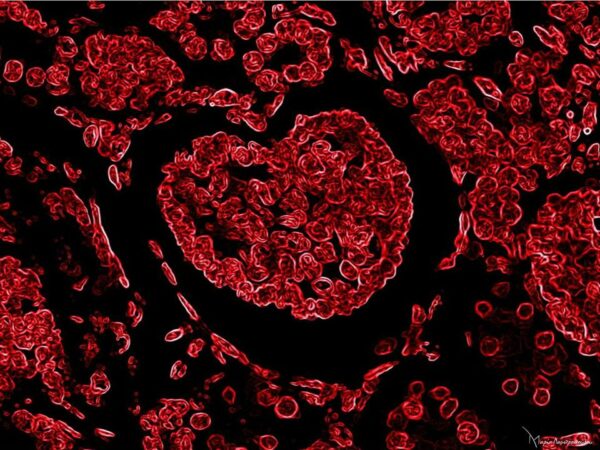
Cover image on Nephrology Dialysis Transplantation (NDT) journal's cover, in the February 2025 issue. Oxford University Press. Kidney histological section in which a glomerulus forming the most iconic symbol of love. The cover image is dedicated to month of love and Valentine's Day. (H&E staining, 200X negative colors); from the "Micro Life Art" collection by Maria Lambropoulou (https://academic.oup.com/ndt/issue/40/2). ©Maria Lambropoulou.
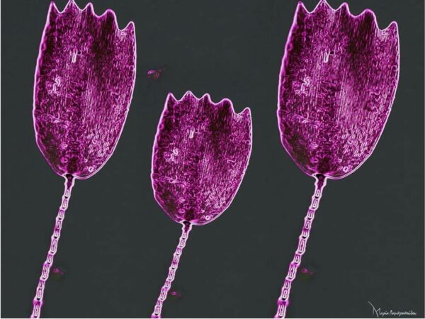
“Toulips”, 2021: Entrapped part of a butterfly wing, during histological section procedure. Light microscope Nikon Eclipse 50i; camera Nikon Digital Sight DS-L1 (Nikon Corporation, Japan); magnification x200. ©Maria Lambropoulou.
“Wild flowers”, 2021: Human Mesothelial Cells, cytological stain Papanikolaou (PAP). Light microscope Nikon Eclipse 50i; camera Nikon Digital Sight DS-L1 (Nikon Corporation, Japan); magnification x400. ©Maria Lambropoulou.
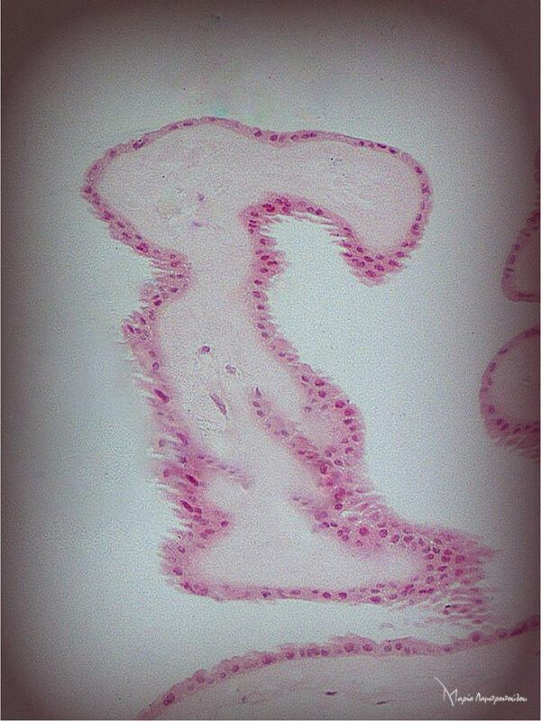
“Winged Victory of Samothrace”, 2018: Tissue section of fetal membranes, Hematoxylin & Eosin staining. Light microscope Nikon Eclipse 50i; camera Nikon Digital Sight DS-L1 (Nikon Corporation, Japan); magnification x100. ©Maria Lambropoulou
“Foresta Rosa”, 2019: Glue and color mixed out, over the edge of section during histological section procedure. Light microscope Nikon Eclipse 50i; camera Nikon Digital Sight DS-L1 (Nikon Corporation, Japan); magnification x100. ©Maria Lambropoulou.
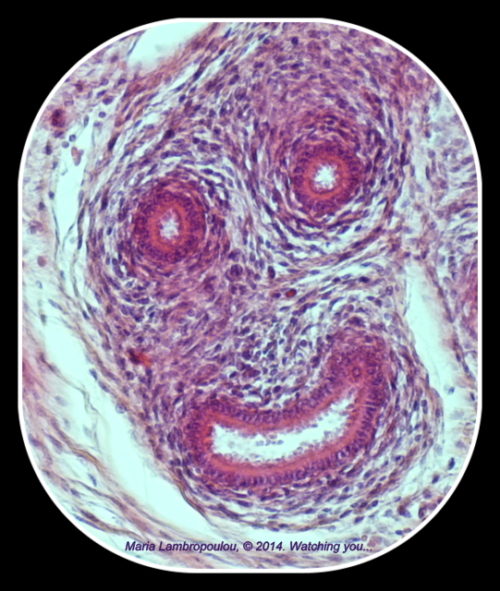
"Watching You!!!" Fetal epididymis section, H&E. Light microscope Nikon Eclipse 50i; camera Nikon Digital Sight DS-L1 (Nikon Corporation, Japan); magnification x200. ©Maria Lambropoulou
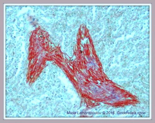
"Cinderella's Little Glass Slipper". Spleen's trabeculae, Masson's Trichrome Staining. Light microscope Nikon Eclipse 50i; camera Nikon Digital Sight DS-L1 (Nikon Corporation, Japan); magnification x200. ©Maria Lambropoulou
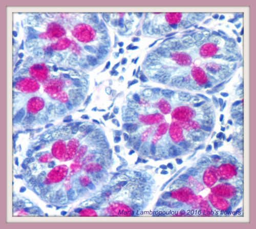
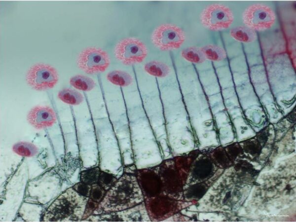
"The Flowers of Our Inner World". Intestinal glands, PAS staining. Light microscope Nikon Eclipse 50i; camera Nikon Digital Sight DS-L1 (Nikon Corporation, Japan); magnification x200. ©Maria Lambropoulou
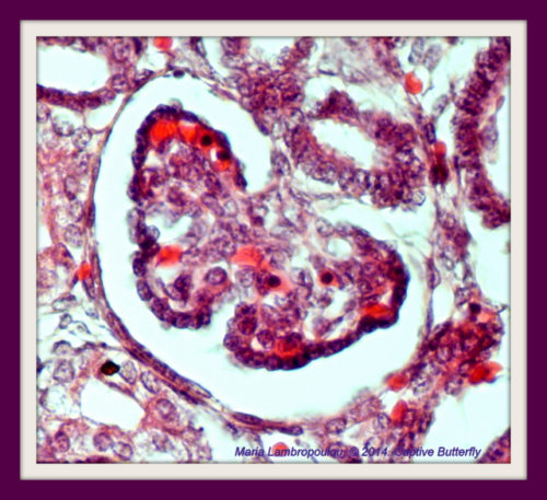
"Captive Butterfly, Symbol of Freedom and Transformation". Renal corpuscle, Hematoxylin & Eosin staining. Light microscope Nikon Eclipse 50i; camera Nikon Digital Sight DS-L1 (Nikon Corporation, Japan); magnification x200. ©Maria Lambropoulou
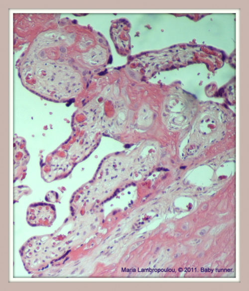
«Baby Runner». Placenta’s section, Hematoxylin & Eosin staining. Light microscope Nikon Eclipse 50i; camera Nikon Digital Sight DS-L1 (Nikon Corporation, Japan); magnification x200. ©Maria Lambropoulou
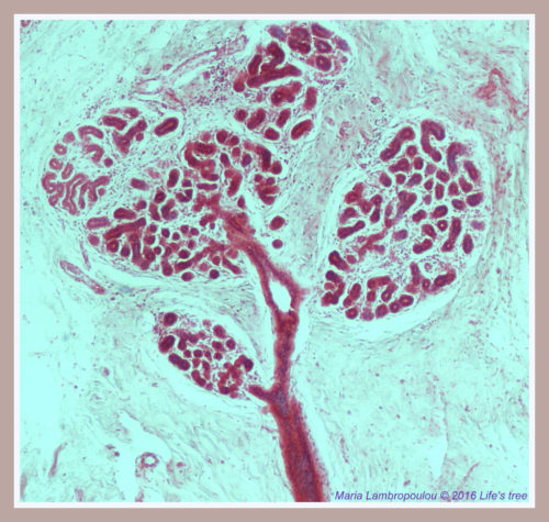
«Life’s Tree». Breast tissue section, Hematoxylin & Eosin staining. Light microscope Nikon Eclipse 50i; camera Nikon Digital Sight DS-L1 (Nikon Corporation, Japan); magnification x200. ©Maria Lambropoulou
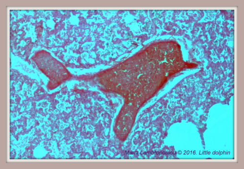
"Pink Dolphin: a beautiful endangered species" Vessel section of fetal lung, H&E staining. Light microscope Nikon Eclipse 50i; camera Nikon Digital Sight DS-L1 (Nikon Corporation, Japan); magnification x200. ©Maria Lambropoulou
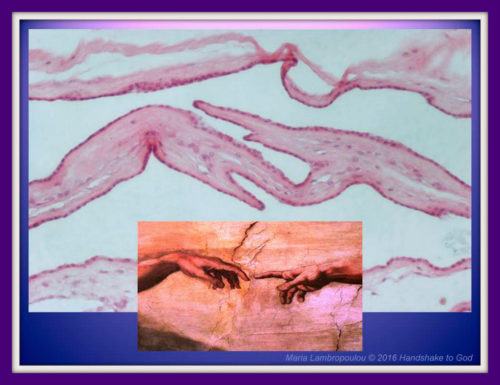
«Handshake to God». Tissue section of fetal membranes, Hematoxylin & Eosin staining. Light microscope Nikon Eclipse 50i; camera Nikon Digital Sight DS-L1 (Nikon Corporation, Japan); magnification x200. ©Maria Lambropoulou
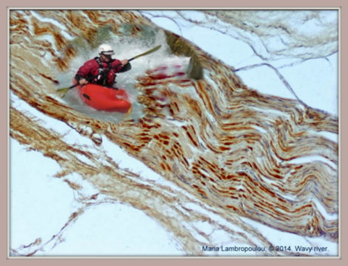
«Rower on Wavy River”. Peripheral nerve section, Neuron Specific Enolase (NSE), immunohistochemical staining. Light microscope Nikon Eclipse 50i; camera Nikon Digital Sight DS-L1 (Nikon Corporation, Japan); magnification x200. ©Maria Lambropoulou
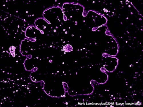
«Space imagination». Air bubbles and glue between coverslip and slide (negative colors). Light microscope Nikon Eclipse 50i; camera Nikon Digital Sight DS-L1 (Nikon Corporation, Japan); magnification x200. ©Maria Lambropoulou
“Medallion of roses”, 2020: Kidney tissue section, renal corpuscle, Hematoxylin & Eosin staining, negative colors. Light microscope Nikon Eclipse 50i; camera Nikon Digital Sight DS-L1 (Nikon Corporation, Japan); magnification x200. ©Maria Lambropoulou.
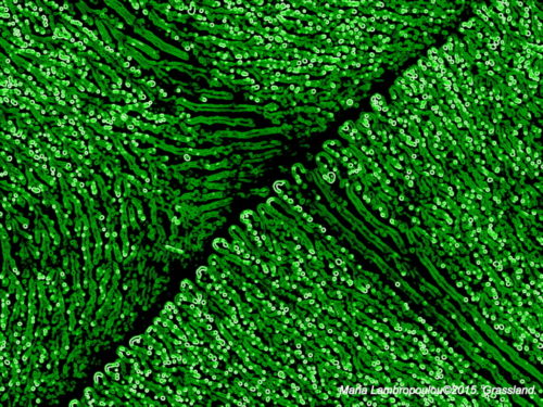
«Grassland». Excess of glue on slide’s surface (negative colors). Light microscope Nikon Eclipse 50i; camera Nikon Digital Sight DS-L1 (Nikon Corporation, Japan); magnification x200. First prize in "Art Path", Belgrade, September 2015 in the frame of European Congress of Pathology. ©Maria Lambropoulou
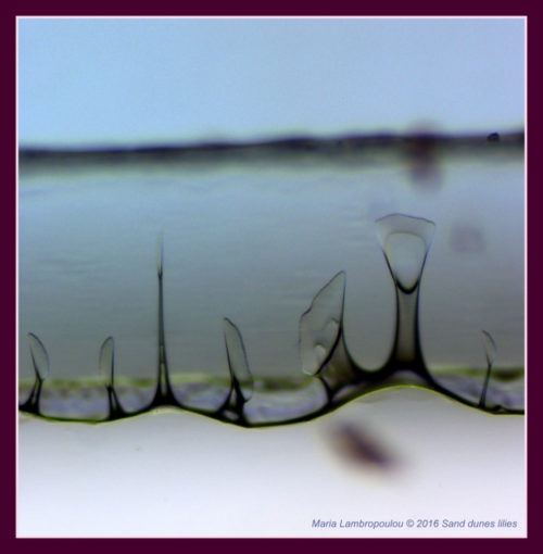
«Sand Dunes Lilies». Air bubbles and glue between coverslip and slide. Light microscope Nikon Eclipse 50i; camera Nikon Digital Sight DS-L1 (Nikon Corporation, Japan); magnification x200. ©Maria Lambropoulou
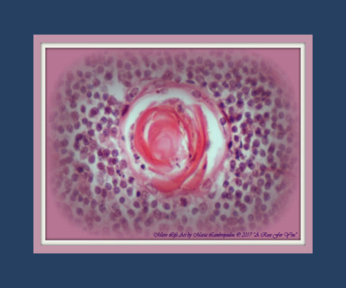
“A rose for you”, 2017: Thymus gland section, Hassall’s corpuscle. Hematoxylin & Eosin staining. Light microscope Nikon Eclipse 50i; camera Nikon Digital Sight DS-L1 (Nikon Corporation, Japan); magnification x200. ©Maria Lambropoulou
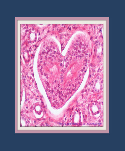
“Double heart”, 2014: Kidney’s section, renal tubules. Hematoxylin & Eosin staining. Light microscope Nikon Eclipse 50i; camera Nikon Digital Sight DS-L1 (Nikon Corporation, Japan); magnification x200. ©Maria Lambropoulou
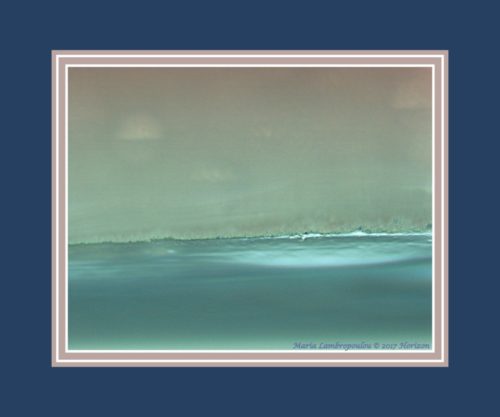
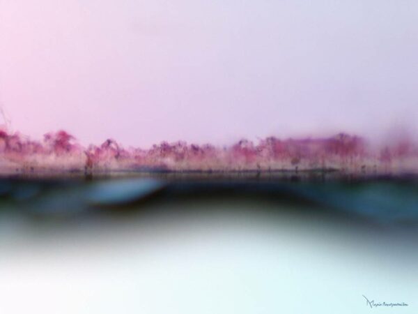
“Horizon”, 2017: Glue and entrapped dust particles, under the coverslip, during histological section procedure. Light microscope Nikon Eclipse 50i; camera Nikon Digital Sight DS-L1 (Nikon Corporation, Japan); magnification x200. ©Maria Lambropoulou
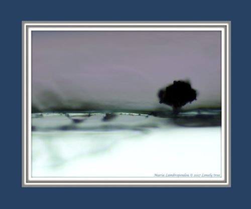
“Lonely Tree , 2017: Glue and entrapped dust particles, under the coverslip, during histological section procedure. Light microscope Nikon Eclipse 50i; camera Nikon Digital Sight DS-L1 (Nikon Corporation, Japan); magnification x200. ©Maria Lambropoulou

Maria Lambropoulou
